The study of non-living and living matter exposed to radiations addresses both fundamental and applied questions in the fields of health, materials and energy. The interaction of ionizing radiations with matter can induce physical, chemical or biological transformations that occur over an exceptionally wide timescale, from the emission of prompt gamma rays of a few 10-20 seconds after exposure to long-term biological consequences. These studies require experimental developments and suitable modeling tools. In this key area, teams of physicists, chemists and biologists work together to see this research through to a successful conclusion.
The skateholders involved are the GIP ARRONAX, the CRCI2NA and Subatech, but also external laboratories within the framework of national and international collaborations. The themes addressed concern:
- Nuclear medicine
- Production of innovative radionuclides: measurements of cross-sections and targetry
- Radiolysis of radiopharmaceutical products
- Materials and energy
- Radiolysis of aqueous mediums
- Speciation of radionuclides under radiation for the fuel cycle
- Non-destructive analysis and inspections
- Ion Beam Analysis (IBA)
- Non-destructive testing (NDT)
- Radiobiology
- Cellular culture and biological monitoring (conventional techniques and real-time microscopy)
- Alpha-beam radiation of cells (vertical or horizontal): inspection of the physical dose, production and effects of radical species, pulsed radiolysis study
- Studies of the effects of strong dose rates (flash proton therapy) and the chemical species produced
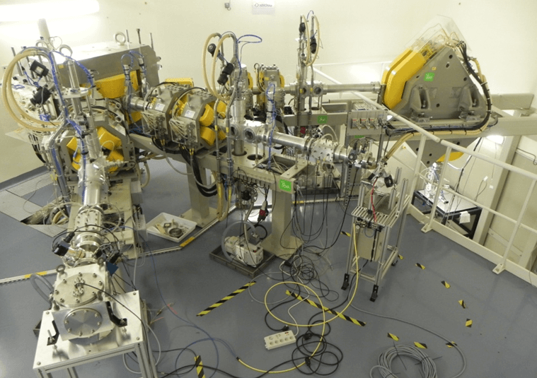
This research relies on state-of-the-art equipment: electronic microscope, electron paramagnetic resonance spectrometer, X-ray tomograph, timelapse microscope, etc. (see “Our equipment” section) and a broad range of available particles and energies: protons, deuterons and alpha particles (produced by the Arronax cyclotron, up to 70 MeV); neutrons (neutronic activator); gamma and X photons (generators and irradiators).
Innovative methods and tools are also used and developed:
- Beam
- 3 dedicated radiation lines, including a vertical one
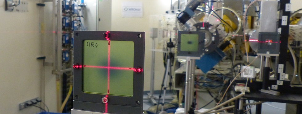
- Development of beam diagnostic tools
- Development of pulsed beams
- Dosimetry
- In-line dosimetry, non-invasive, physical and chemical
- Development of detectors and related methods
- Cellular culture
- Ability to work in a sterile medium
- Incubators, inverted microscope, freezer at -80°C
- Fluorescence time-lapse microscopy
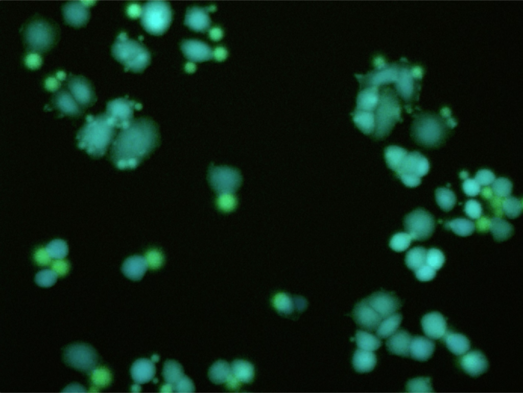
View of breast cancer cells in the presence of a fluorescent tracer
after radiation by At-211
our publications
Methodology for small animals targeted irradiations at conventional and ultra-high dose rates 65 MeV proton beam, Evin M, Koumeir C, Bongrand A, Delpon G, Haddad F, Mouchard Q, et al., Physica Medica., 1 avr 2024;120:103332. https://doi.org/10.1016/j.ejmp.2024.103332
Proton Irradiations at Ultra-High Dose Rate vs. Conventional Dose Rate: Strong Impact on Hydrogen Peroxide Yield, Blain G, Vandenborre J, Villoing D, Fiegel V, Fois GR, Haddad F, et al., Radiation Research, June 8, 2022.
https://doi.org/10.1667/RADE-22-00021.1How radiolysis impacts astatine speciation?, Ghalei M, Mahdi Khoshouei P, Vandenborre J, Guerard F, Blain G, Zarei M, et al., Radiation Physics and Chemistry. sept 2022;198:110224.
https://doi.org/10.1016/j.radphyschem.2022.110224
our news
Lunar Radon detectors under test at Arronax
23.05.2022