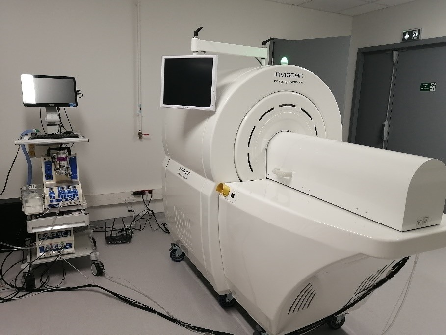
IRIS PET/CT is dedicated to preclinical in-vivo research on small animals such as mice and rats. Our bimodal equipment is coupled with a gas anesthesia system with monitoring.
Characteristics
TEP :
- Sensitivity: > 9% [250 – 750 keV]
- Spatial resolution = 1.1 mm (MLEM)
- Axial FOV: > 94 mm (Recon FOV = 102.6 mm)
- Trans-axial FOV: ~ 81 mm
- Energy resolution: < 13%
- Timing resolution = 1.8 ns
CT :
- Detector imaging area: > 11.4 x 14.5 cm2
- Scan time: < 7.3 s (ultra-fast mode), 20 s (speed mode), 1 min (high resolution scan)
- Low dose: < 6.5 mGy
- Resolution: 73μm @ 10% MTF
- Minimal voxel resolution: < 30μm
- Axial FOV: > 90 mm
- Dynamic 4D acquisition
Principles
PET imaging is based on the detection of photons produced during the annihilation of a positron with an electron. Positrons are emitted by artificial radioactive isotopes with short half-lives such as fluorine-18 (half-life around 2 hours) that are introduced into a molecule of interest to obtain images of the chemistry of living organisms: tissue perfusion, metabolism (glucose, oxygen), receptor density and affinity, enzymatic activity, expression of reporter genes, or biodistribution of therapeutic molecules. The strong points of PET are its very high sensitivity, excellent signal-to-noise ratio, the quantitative and intrinsically tomographic nature of the information, a detection depth of several tens of centimeters, the availability of a large number of radiotracers, and the absence of toxicity due to the small amount of substance used and the rapid elimination of radioactivity. The PET image is associated with the X-ray scanner for anatomical location.
| Identity card | ||
| Name | PET/CT IRIS | |
| Manufacturer | INVISCAN | |
| ID number (internal ref.) | ||
| Place | CIMA | |
| Owner | Université de Nantes | |
| Type of equipment | PET/CT | |
| Operation | ||
| Availability of the equipment | Available | |
| Apparatus in a controlled zone | Yes | |
| Restrictions on use | Mandatory supervision | |
| Technical correspondent | Contact us | |