General presentation
This equipment is a scanning electron microscope which makes it possible to produce 2D images. It is coupled with EDS to determine and quantify chemical elements. It has been installed at GIP ARRONAX since 2015.
Principle
Surface micro-analysis is regularly used to characterize the surface state of a sample. The SEM JEOL JSM 7100F installed at GIP ARRONAX has enabled the R&D group to study the metallic surface state in details by using secondary electrons 1. to make images, and backscattered electrons and 2. to check the homogeneity of the chemical elements, and surface relief. EDS micro-analysis coupled with the SEM is used to identify the chemical elements present on the surface of a sample.
Exemple of use
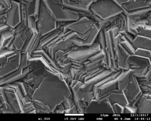
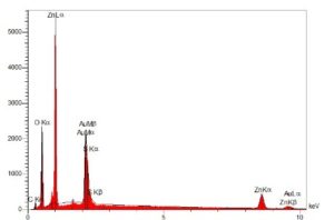
The images below show a zinc deposit made by electrodeposition (Zn) on a gold (Au) substrate: visible deposit (top left); through the SEM (top right); EDS spectrum (lower). These images show that defects are absent: these deposits do not contain impurities (the Au peaks come from the substrate) and the crystalline structure of the Zn deposit is hexagonal with a three-dimensional stack. The zinc deposit is suited for irradiation for the production of copper-67 for radiopharmaceutical purposes.
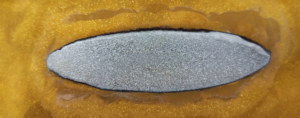
The shape of the deposit is elliptical, with the following dimensions: 1.1 cm of semi-minor axis, 4 cm of semi-major axis, 149 µm and thickness.
| Identity | |
| Name of the equipment | MEB JEOL JSM 7100F |
| Manufacturer | JEOL |
| Location | GIP ARRONAX |
| Name of the owner institution | GIP ARRONAX |
| Type of equipment | Scanning Electron Microscope |
| Operation | ||
| Availability | Available | |
| Equipment in restricted area | Yes | |
| Constraints of use | Mandatory training | |
| Technical contact | Contact us | |